

Highlightfeatures
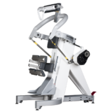
CON-TREX® TP 500
- hochpräziser Isokinetik-Modus
- Ballistik-Modus
- aktive Kompensation der Schwerkraft
- einzigartige Bewegungsabfolgen
Allgemeine Features
- Abtastrate von 4000 Hz für höchste Präzision
- spielfreier Antriebsstrang
- EMG-Synchronisation
- dreistufiges Sicherheitskonzept
- individuell anpassbare Reports

Durch ein innovatives, ballistisches Regelungsverhalten werden mittels Vorausberechnung der zu erwartenden Bewegung höhere Beschleunigungen und damit eine schnellere Bewegung ermöglicht. Es resultiert eine deutliche Reduktion des Einflusses der Trägheitsmomente. Im ballistischen Modus können höhere Bewegungsgeschwindigkeiten bzw. eine definierte Geschwindigkeit kann länger isokinetisch ausgeführt werden. Der Ballistik-Modus optimiert Training und Diagnostik mit funktionellen und realitätsnahen Bewegungen und Belastungen.

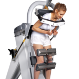
Patienten mit geringer Kraft sind häufig nicht in der Lage, einzelne Körpersegmente ohne Unterstützung zu bewegen. Es braucht daher über die ganze Bewegungsamplitude hinweg eine aktive Kompensation dieser statischen Gewichtseinflüsse. Während der Bewegungsausführung kann das Dynamometer die externen Kräfte laufend reduzieren oder sogar vollständig ausgleichen; für den Patienten resultiert damit eine «schwerelose» Situation, in der sich jede Bewegung mit minimalstem Kraftaufwand ausführen lässt.

Die äußerst hohe Abtastrate von 4000 Hz stellt die extrem präzise Regelung des Dynamometers sicher und reduziert den unerwünschten Jitter (zeitliches Taktzittern in der Regelungstechnik) auf ein nicht mehr wahrnehmbares Minimum. Durch den spielfreien Antriebsstrang ist während der Bewegungsumkehr kein Spiel spürbar, was vor allem für hochdynamische Bewegungen (wie zum Beispiel Werfen) relevant ist. Dies ermöglicht auch die Ausübung einzigartiger Bewegungsabfolgen wie zum Beispiel die Kombination der kontinuierlich-passiven Bewegung (CPM) mit konzentrischer oder exzentrischer Belastung.

Der konstante Offset von 12 Millisekunden bei voller Abtastrate (4000 Hz) ermöglicht eine präzise, einfache und zuverlässige Synchronisierung der Signale.

Die stufenlos einstellbaren mechanischen Bewegungsstops limitieren direkt am Dynamometer den Bewegungsbereich. Zusätzlich wird die Position der Bewegungsstops vom System erkannt und laufend mit dem ausgewählten Movement Pattern abgeglichen. Somit wird ein Überschreiten der Bewegungsamplitude durch die Software redundant verhindert.

Bei CON-TREX®-Systemen wird der Dynamometer zum Patienten bewegt. Der flexible Dynamometer-Kopf erlaubt die präzise und äußerst nahe Einstellung der Drehachse des Dynamometers in Bezug auf die Gelenksachse.
Das breite Adapter-Sortiment ist aus hochfestem nichtrostenden Edelstahl gefertigt und vereinigt eine maximale Verwindungssteifigkeit mit geringstmöglichem Massenträgheitsmoment. Die Verstellung erfolgt stufenlos und zeitsparend durch einen ausgeklügelten und zuverlässigen Klemmmechanismus.
Individuell anpassbare Reports mit vielfältigen Darstellungsmöglichkeiten und Diagrammen und Exportfunktion als PDF-Dokument. Der Export der Messdaten per ASCII-File erlaubt die Weiterverarbeitung in anderen Programmen.

human kinetics Software
Die human kinetics Software ermöglicht die einfache Trennung von Datenbanken, wodurch in einer Einrichtung verschiedene interne Abteilungen oder wissenschaftliche Bereiche unabhängig voneinander arbeiten können. Die Darstellung der zahlreichen Reportdesigns lässt sich individuell anpassen. Der Export in weitere Datenverarbeitungsprogramme geschieht einfach via ASCII-Schnittstelle. Für wissenschaftlichen Arbeiten ist eine Anonymisierung der Daten problemlos möglich.
In der detaillierten Online-Hilfe werden direkte Hinweise zum aktuellen Menüpunkt oder der gerade ausgeführten Bedienung mit Bildern und Grafiken gegeben. Vor allem die detaillierten Angaben zu den zahlreichen Bewegungsausführungen verdeutlichen sowohl die Positionierung des Probanden/Patienten wie auch die Verwendung der benötigten Adapter.
Therapieinformationen
CON-TREX® kommt in der frühen diagnostischen und präventiven Therapie bei Verletzungen des Bewegungsapparates in der ambulanten Rehabilitation und in der Klinik zum Einsatz. Ferner wird es in der wissenschaftlichen Forschung und Leistungsoptimierung genutzt und ermöglicht die sorgfältige spezifische Problemauswertung und somit hocheffizientes Training von Hochleistungssportlern.
CON-TREX®-Geräte eignen sich auf Grund ihrer hohen Präzision zur Messung und Analyse, und sind deshalb besonders für den wissenschaftlichen Einsatz geeignet. Bei Training und Therapie zielen sie auf eine Verbesserung muskulärer (im Kraft- wie auch im Kraftausdauerbereich) und sensomotorischer Fähigkeiten ab. CON-TREX® ist Dank seiner vielseitigen Messmöglichkeiten und der attraktiven Übungs-Software für folgende Anwendungen hervorragend geeignet:
CON-TREX® ermöglicht frühe Diagnostik und Prävention von Schädigungen oder Verletzungen des Bewegungsapparates in der ambulanten Rehabilitation und im klinischen Einsatz.
Diagnose und Rehabilitation muskuloskelettaler Defizite
Muskuläre Dysbalancen können den optimalen Bewegungsablauf stören und sich gelenkschädigend auswirken, bzw. sportartspezifisch erwünscht oder gefordert sein. CON-TREX® hilft diese zu erfassen, aufzudecken und zu analysieren. Zusätzlich können die CON-TREX®-Geräte bei der Beseitigung von muskulären Dysbalancen effizient eingesetzt werden. Ein besonderer Vorteil besteht darin, dass die getestete Bewegung gleichzeitig trainiert werden kann.
Gelenkersatz
Vor allem im Bereich der geriatrischen Rehabilitation kommen die CON-TREX®-Geräte nach künstlichem Gelenkersatz zum Einsatz. Patienten können selbst bei sehr gering vorhandener Muskelkraft aktiv und in einer sinnvollen Bewegungsgeschwindigkeit trainieren und ihre Muskelkraft verbessern. Dadurch wird der Kraftverlust so gering wie möglich gehalten und die Beweglichkeit der Gelenke bleibt erhalten bzw. wird verbessert.
Bei neurologisch bedingten Leistungsminderungen, wie beispielsweise nach Hirnverletzungen oder Schlaganfall, steht die Wiedererlangung von Koordination und Kontrolle über die Muskelarbeit im Zentrum rehabilitativer Arbeit. Die Deutsche Gesellschaft für Neurologie fordert eine frühfunktionelle Mobilisation des Patienten nach Schlaganfall. CON-TREX® eignet sich hierzu durch die Übungs- und Trainingsmöglichkeit im kontinuierlichen passiven Bewegungs-Modus: Die betroffene Extremität des Patienten wird durch CON-TREX® bewegt und der Patient kann gleichzeitig versuchen, die Extremität selbst anzusteuern und zu bewegen. CON-TREX® visualisiert zeitgleich die aktuelle Leistung des Patienten, d.h. das Training wird am Bildschirm in Echtzeit verfolgt und selbst kleinste Fortschritte sind unmittelbar nachzuvollziehen. Dies trägt zur Motivation des Patienten bei, die Wirksamkeit der Rehabilitation durch seine aktive Mitarbeit zu steigern. Mit «klassischen» Trainingsverfahren lässt sich dies nur eingeschränkt erreichen. Bio-Feedback-Training, insbesondere unter submaximaler Belastung, ermöglicht nicht nur die effiziente Korrektur von muskulären Defiziten, sondern ist auch eine ausgezeichnete Methode zur Verbesserung der koordinativen Fähigkeiten.
CON-TREX®-Geräte finden ihren Einsatz im Leistungssport vor allem in der objektiven Verfassungsbeurteilung und Optimierung des Trainingsverlaufs von Leistungs- und Hochleistungssportlern. Mit verschiedenen Krafttests, die in regelmäßigen Abständen durchgeführt werden, bekommen Trainer wie auch Sportler eine exakte Rückmeldung über die Effektivität ihres Trainings. Im Rahmen von Bewegungsanalysen zur Optimierung des sportartspezifischen Bewegungsablaufes können darüber hinaus mit kombinierten EMG-Auswertungen exakte Problemanalysen erstellt werden. In der Rehabilitation von Leistungssportlern nach Verletzungen ermöglichen CON-TREX®-Geräte ein hocheffizientes Training und tragen dadurch zu einer sinnvollen Nutzung der Verletzungszeit bei.
Dank der hohen Messgenauigkeit der CON-TREX®-Geräte ist eine objektive Auswertung jedes Patienten mit höchstmöglicher Validität gegeben. CON-TREX® archiviert alle relevanten System-Parameter, die für eine wissenschaftliche Auswertung sinnvoll sein könnten. Zusätzlich sichert der einzigartige Ballistik-Modus einen reibungslosen Bewegungs- und Messablauf. Die aktive Schwerkraft-Kompensation ermöglicht eine absolute und relative Betrachtung der Werte. Für die Verwendung in Wissenschaft und Forschung setzen die CON-TREX®-Geräte in Sachen Messgenauigkeit sowie Reproduzierbarkeit der erhobenen Parameter einen bisher unerreicht hohen Standard.
